- Homepage
- Key Information
- Students
- Staff
- PGR
- Health and Safety
- Computer Support
- National Student Survey (NSS)
- Intranet Help
Prof Frank Vollmer
Research
I am pursuing a multi-disciplinary research initiative in Molecular, Nano- and Quantum Sensors and Systems that is unique in the UK (and the world) and that brings together the research streams of nanophotonics, nanoplasmonics, quantum optics, molecular mechanics (molecular machines, synthetic bio) and in the future, also molecular electronics and neuroscience. This new pan-disciplinary area, I believe, will be a very large and upcoming research playground at the cross-roads of cutting edge experimental and theoretical sciences; there will be applications in health, nanotechnology, metrology, environment, security, and astronomy; it touches on core subjects in physics, quantum optics, optics, biophysics, engineering, molecular mechanics and biochemistry.
Vollmerlab web page: https://www.vollmerlab.com/
Review: Whispering-Gallery-Mode Sensors in Physical and Biological Sensing https://rdcu.be/cCURa
PhD studentships and postdoc positions are available in the Vollmer Lab:
Postdoctoral positions
The project will develop chemical control at the nanoscale for single molecule sensing of molecular machines:
https://jobs.exeter.ac.uk/hrpr_webrecruitment/wrd/run/ETREC107GF.open?VACANCY_ID=875598O4ZX&WVID=3817591jNg&LANG=USA
we are also looking for a theorist, advert on nature careers
more PhD/postdoc job posting for mygroup on on nature careers, physics today, researchgate and exeter jobs!
Observing the Motions of Nano-Machines
Have you ever wondered about how our bodies might work at the nanoscale, a scale at which we are composed of individual biomolecules? Where individual biomolecules such as enzymes take on the role of molecular machines, and where parts of a protein move like the pistons of an engine? How can one observe and analyse such intricate systems?
My research aims to address these questions. My laboratory develops optical techniques to directly visualise living machinery. We are interested in looking at biological nanomachines, such as motor proteins and enzymes, as they function and while disturbing them as little as possible.
Our micro-optical devices and spectrometers have important applications in health, environment, security, and astronomy.
With our optical sensors, processes at the nanoscale can be studied with great precision. The observation of single atomic ions is just a first step towards exploring the ultimate limits of detection. By implementing advanced metrology and techniques from laser interferometry and atomic optics, further breakthroughs in nanoscale precision measurements are anticipated. Techniques from quantum optics combined with novel materials may enable yet unexplored sensing strategies.
Taking Single-Molecule Detection to the Limit
In 2014, we built the world’s most sensitive optical device capable of detecting single-molecules without chemical alteration (1). The platform is based on optical microcavities, approximately 100 um diameter glass microspheres that are used as optical sensors. Optical resonances, so-called Whispering gallery modes (WGMs), were excited in the microspheres for the label-free detection of biomolecules(2, 3). These microsphere sensors were used to detect single DNA molecules and their interaction kinetics(1).
In 2015-2016, we further advanced these optical sensors, improved upon the detection limit and time resolution. Advanced experimental capabilities of these sensors led to the publication of seminal works, on the sensing of single atomic ions in solution(4), the detection of various single-molecule surface reactions from low to high affinity(5), and on unprecedented nanosecond time resolution for label-free single-molecule studies(6). These demonstrations consolidate our optical technique as one of the most sensitive tools for label-free single molecule studies. They establish a biosensor technology that can detect and analyse the intricate dynamics of single biomolecules. In most recent works we demonstrate such capability, with the first label-free optical technique capable of observing enzymatic interactions and associated conformational changes on the single molecule level(7).
The future of our micro-optical sensors is outlined in a recent roadmap(8). To summarize, our optical sensors can help us understand how our bodies work at the nanoscale, where individual biomolecules such as enzymes take on the role of nano-machines, and where parts of a protein move similar to the pistons of an engine. Without the need of a label, our sensors will provide a universal tool for the unabated exploration of structural dynamics and shape-changes in individual proteins. Our optical devices can furthermore harness the extreme speed, selectivity and specificity of the biological nanoworld. With further technological advances already in the pipeline, our sensors will benchmark nanoscale metrology. Those with ultimate sensitivity will provide a tool for uncovering novel physical phenoma at the nanoscale.
In a second line of experiments, we have established experiments with two-dimensional photonic crystal structures. These structures can provide optical sensing devices that are highly suitable for on-chip biosensing applications. In photonic crystals, light is confined in engineered defect cavities or by means of Anderson Localization - as we have uncovered in our previous research. Recently, we have focused on establishing a photonic crystal biosensing platform by leveraging Anderson Localization and free-space coupling with polarization-tailored beams. With such a platform, we realise various on-chip single-molecule biosensing applications.
The following sections highlight the individual research projects.
Single-Ion Sensing
Plasmonic nanoparticles (NPs) have been used for a wide range of applications, such as spectroscopy, high-resolution imaging, medical therapy, nonlinear optics and photocatalysis. Such widespread utility is attributable to the diverse range of NP shapes and sizes available and the corresponding tunability of their optical properties. The highly localized fields near the surface of such particles, in addition to their small mode volumes, make them especially suitable for sensing or even capable of label-free single-molecule detection.
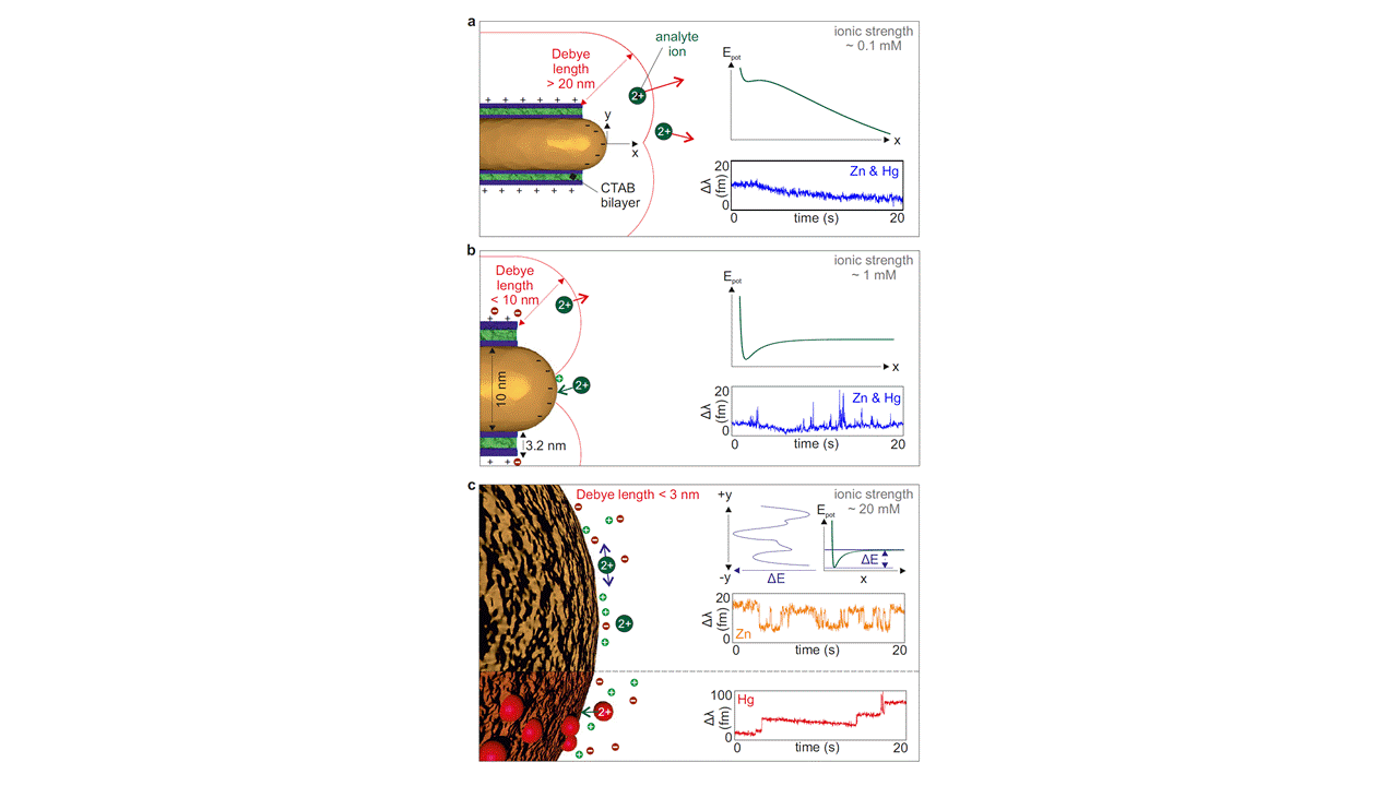
In 2016, we have shown that the sensing capability based on plasmonic effects in nanpoarticles can be boosted with optical microcavities for detecting even the smallest chemical species in aqueous solution, single atomic ions(4), see Figure 1.We achieved single-molecule sensitivity by resonantly coupling nanorods (NRs) to optical microcavities.
This is a landmark-breakthrough in the area of optical sensing technology as the observation of single atomic species has thus far been restricted to investigations in vacuum, typically using atom traps. Our interferometric technique based on plasmon-enhanced whispering gallery mode sensors brings these investigations to the solution phase at room temperature. Our study lays the foundation for future all optical investigations of atomic processes at nanoparticle sensor surfaces in the liquid phase, such as investigation of the optical interaction of plasmons with diffusing and bonded ions, analysis of the physical chemistry of atomic processes at nanoparticle surfaces, investigations of surface potentials with single ion probes, and environmental sensing of hazardous chemicals such as mercury, In a related experiment, we have demonstrated the highly specific environmental sensing of mercury ions, using a DNA strand displacement reaction integrated with a WGM sensor(9).

Figure 1: Optical observation of single atomic ions Zn2+ and Hg2+ interacting with plasmonic nanorods in solution. The detection signal has been boosted with optical microcavities. Three different interaction regimes (a)-(c) were observed, which depend on the ionic strength of the solution. Adapted from (4).
In-Situ Observation of Single-Molecule Surface Reactions from low to high Affinity
Nanoparticles have become ubiquitous part of nanotechnology and progress to far more mature applications including medical therapy, drug delivery, biological sensing and imaging. The implementation of nanoparticles for such real life applications requires the development of functional surface modifications to meet the specific requirements set by the application itself. Many researcher have therefore put their efforts together to develop protocols to modify the surfaces of diverse nanomaterials. These efforts, however, are somewhat hindered as most metrologies employed to test the modifications are limited to reaction end products thus resulting in time intensive trial and error phases in the early development stages.
In 2016, we have developed a completely new approach to study surface-based interactions by utilizing WGM microcavity sensors(5). This all optical approach resolves transient as well as permanent interactions of single ligand molecules at the nanoparticle interface in situ, Figure 2. Reaction kinetics can thus be monitored over a broad range of affinities and especially under conditions where no reaction product is formed on average. This enables the real time observation and characterization of reactions during the entire procedure of a bottom-up surface modification, ranging from the deposition of ligands to the confirmation of their functionality. This technique will be a powerful tool, not only for extending the fundamental knowledge of molecular interactions occurring on nanomaterial interfaces, but also for the rapid development of new functionalization protocols.
Figure 2: Top: Illustration of surface reactions with low and high affinity. Bottom: The corresponding single-molecule WGM sensor signals. Spikes indicate a low affinity, steps indicate a high-affinity single-molecule reaction. Adapted from (5).
Nanosecond Time-Resolution for WGM Sensors
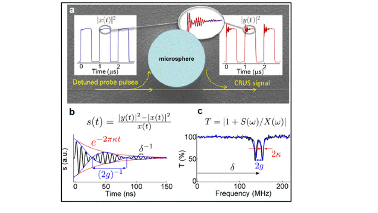
Currently, most WGM sensors operate with time resolutions of milliseconds to seconds, depending on the operational requirements, whereas these detection speeds are mostly not limited by the sensor system itself but by the method employed to read out the resonator’s spectral response. In scenarios where the swept wavelength modality is used to obtain the WGM spectra, the laser’s maximum scanning speed is the major limiting factor. In principle, the intrinsic time resolution of the system is set by the decay rate of the cavity, which is typically below one microsecond. Recent experimental works from in collaboration with S. Rosenblum et al.(6)achieved the detection of submicrosecond optomechanical vibrations of microtoroids by a cavity ring-up spectroscopy (CRUS), see Figure 3. In this system, a far-detuned (with respect to the WGM) probe laser is abruptly switched on creating a broader spectrum around the laser’s center frequency, where parts of the spectrum is coupled into the resonator. Afterwards, the probe laser reaches cw operation and is thus not coupled to the WGM any longer, while a fraction of the previously excited modes is continuously coupled out of the cavity, where it interferes with the probe laser, thus creating a ring down interferogram, from which the WGM’s lineshape and position can be obtained via Fourier transformation. This technique, therefore, is very similar to the cavity ring down spectroscopy (CRDS), where the decaying emission intensity is measured after the excitation laser is switched off in order to obtain the cavity lifetime. Unlike CRDS, however, CRUS provides the complete spectral information including centre frequency and linewidth of the WGM Mode (including mode split), which thus can be used for monitoring spectral shifts induced by molecules/particles.

Figure 3: Cavity ring-up spectroscopy for ultrafast sensing with optical microresonators. Adapted from (6).
Identifying a novel biomarker for apoptosis and cell death
We have teamed-up with the Institute for Biochemistry and the University Hospital in Erlangen to investigate the response of cells to toxins using optical microcavities. The response of cells to toxins is commonly investigated using biochemical tools for detecting intracellular markers for cell death, such as caspase proteins. This requires the introduction of labels by the permeabilization or complete lysis of cells. In our study(10), we have introduced a non-invasive optical tool for monitoring a caspase protein in the extracellular medium. The tool is based on highly sensitive optical micro-devices, referred to as WGM biosensors (WGMB’s). WGMB’s were functionalized with antibodies for the specific and label-free detection of procaspase-3 released from human cells after introducing toxins. These studies provide evidence for procaspase-3 as a novel extracellular biomarker for cell death, with broad applications in cytotoxicity tests. Such tests could be administered on lab-on-chip and organ-on-chip devices, culminating to the first commercial method for non-invasive, rapid, real-time, and extracellular detection of cell death by procaspase-3 markers.
Thermal characterization of Biopolymers
The structure of biopolymers is known to change with temperature, affecting for example the activity of enzymes and the folding of proteins. We have shown that such structural change can be monitored using highly sensitive WGM optical biosensors(11). To achieve this goal, my group has devised a technique to temperature-stabilize the WGM sensor system, by adding glycerol to a WGM glass microsphere placed in an aqueous sensing environment. The temperature stabilized WGM sensor was utilized to track structural changes in biopolymers, by monitoring frequency shifts associated with optical polarizability change. Using this sensing method, structural changes in albumin protein (figure) could be resolved for only one degree in temperature variation. Such highly sensitive detection of structural change can have many important applications: in single molecule studies, for thermodynamic investigations of biological systems, and for monitoring temperature-dependent reaction kinetics.
Stand-Off Biodetection with asymmetric Microsphere Cavities
Instead of using evanescent coupling, direct excitation and detection via free-space optics, where necessary optics can be placed far-away from the sensors, seems promising alternative for WGM sensors. Separation of excitation and detection instruments from the on-chip sensors can simplify the incorporation with microfluidics and may enable in-vivo measurements. Unlike active cavities, however, the free-space coupling of passive high Q cavities is normally an extremely inefficient process. To increase the efficiency to desirable levels, an asymmetric resonator geometry is required. The coupling can then be achieved by focusing a laser beam onto the periphery of cavity where he break in its symmetry occurs, locations which also double up as directional emission points allowing efficient far field read out. We have demonstrated the use of asymmetric microspheres in aqueous medium for the detection of unspecific bulk adsorption of BSA molecules onto the cavity surface(12). In addition, we have visualized the directional emission pattern from the cavity, see Figure 4.
Figure 4: Visualization of the directional emission from an asymmetric microsphere cavity.
Photonic Crystals: Photoluminescence Enhancements
One of the areas of intense research in the field of silicon photonics is the excitation and extraction of photoluminescence (PL). We have demonstrated optimized photonic crystal designs on Silicon on Insulator (SIO) for maximizing light extraction efficiencies supported with extensive computational predictions, see Figure 5. With near optimal design parameters for the PhCs, we reported more than 500-fold increase in PL intensity measured near the band edge of silicon at room temperature, an enhancement by an order of magnitude in respect to what has been previously reported in similar PhC structures with air holes (13). We believe that our investigation has an imperative impact in increasing the device performance of LEDs, solar cells and precision biosensors, providing very robust and generic platforms with optimized predictions.
Figure 5: Photoluminescence enhancement in photonic crystals. (a) Photonic crystal (b) disordered crystal (c) Corresponding photoluminescence spectra. Inset show the simulation results. Adapted from (13).
Photonic Crystals: Free-space Coupling with polarization-tailored Beam
Two dimensional silicon photonic crystals (2D PhC) are often the ideal structure for localizing light in micro-cavities with sub-wavelength modal volumes and quality factors of up to ~2 million. Furthermore, 2D PhCs allow dispersion engineering of waveguide modes with slow light characteristics. The PhC crystal waveguides and -cavities can be excited with a free space beam that is impinging orthogonally onto the PhC slab. This is a particularly attractive geometry for many sensing and light-routing applications.
In a very recent study(14), we have investigated this alternative method for coupling to PhC waveguide modes and for the excitation of microcavities. We have tailored the state of polarization in a near-diffraction limited spot of a focused light beam impinging orthogonally onto a PhC waveguide, Figure 6. We have characterized the efficiency of coupling for various focal electric field density distributions. In conjunction with the disorder present in PhC waveguides, we demonstrate Anderson localization, and excite Anderson-localized microcavities with Q factors ~105.
Figure 6: Excitation of PhC waveguide (PhCWG) modes with polarization-tailored beams.
REFERENCES
1. M. D. Baaske, M. R. Foreman, F. Vollmer, Single-molecule nucleic acid interactions monitored on a label-free microcavity biosensor platform. Nat. Nanotechnol. 9, 933-939 (2014).
2. M. R. Foreman, J. D. Swaim, F. Vollmer, Whispering gallery mode sensors. Advances in optics and photonics 7, 168-240 (2015).
3. M. B. Eugene Kim, Frank Vollmer, Towards next-generation label-free biosensors: recent advances in whispering gallery mode sensors. Lab Chip, volume 17, no. 7, pages 1190-1205, DOI:10.1039/c6lc01595f
4. M. D. Baaske, F. Vollmer, Optical observation of single atomic ions interacting with plasmonic nanorods in aqueous solution. Nature Photonics, volume 10, no. 11, pages 733-739, DOI:10.1038/nphoton.2016.177
5. E. Kim, M. D. Baaske, F. Vollmer, In Situ Observation of Single‐Molecule Surface Reactions from Low to High Affinities. Adv. Mater. 28, 9941-9948 (2016).
6. S. Rosenblum, Y. Lovsky, L. Arazi, F. Vollmer, B. Dayan, Cavity ring-up spectroscopy for ultrafast sensing with optical microresonators. Nature Communications, volume 6, pages 6788-6788, DOI:10.1038/ncomms7788
7. M. B. Eugene Kim, Isabel Schuldes, Peter Wilsch, Frank Vollmer, Label-free optical detection of single enzyme-reactant reactions and associated conformational changes. Science Advances, DOI:10.1126/sciadv.1603044
8. F. Vollmer, A roadmap for whispering gallery mode sensors. to appear in Journal of Optics (2017).
9. F. Wu, Y. Wu, Z. Niu, F. Vollmer, Integrating a DNA Strand Displacement Reaction with a Whispering Gallery Mode Sensor for Label-Free Mercury (II) Ion Detection. Sensors (Basel, Switzerland) 16, (2016).
10. C. Ying-Jen, X. Wei, K. Jochen, V. Frank, Tracking micro-optical resonances for identifying and sensing novel procaspase-3 protein marker released from cell cultures in response to toxins. Nanotechnology 27, 164001 (2016).
11. E. Kim, M. R. Foreman, Martin D. Baaske, F. Vollmer, Thermal characterisation of (bio)polymers with a temperature-stabilised whispering gallery mode microsensor. Appl. Phys. Lett. 106, 161101 (2016).
12. Z. Ballard, M. Baaske, F. Vollmer, Stand-Off Biodetection with Free-Space Coupled Asymmetric Microsphere Cavities. Sensors 15, 8968 (2015).
13. A. Mahdavi et al., Maximizing Photoluminescence Extraction in Silicon Photonic Crystal Slabs. Scientific Reports 6, 25135 (2016).
14. P. R. Ali Mahdavi, Jolly Xavier, Taofiq Paraiso, Peter Banzer, Frank Vollmer, Coupling to Photonic Crystal Waveguide by Polarization Tailored Beam submitted to APL, (2017).
O
